What is skin cancer?
Approximately 5.4 million skin cancers are diagnosed yearly in the United States. By 2040 Melanoma will be the leading type of cancer amongst men. So, if you have been diagnosed or know someone who has, here is what you need to know.
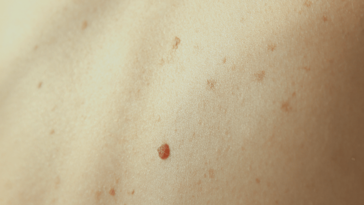
Our body is made of billions of cells that typically are doing basic functions to maintain stability. Skin cancer is a collection of abnormal cells within the skin that are dividing without control and continue to do so until fully treated. In 90% of cases, these changes are caused by accumulated sun damage, but can also be caused by genetics, or even trauma.
What is the most common type of skin cancer?
The most common type of skin cancer is a Basal Cell Carcinoma which is abnormal cell growth within the basal cell layer of the epidermis.

This is the form of skin cancer caused by excessive sun damage to the skin, in particular sunburns. Most of the time it is slow-growing and has an incredibly small chance of traveling elsewhere within the body when caught early. There are many ways to treat a basal cell carcinoma but most dermatologists will take size, type of basal cell carcinoma (aggressive vs. less aggressive types), location, and age into consideration when determining the best treatment option for you.
What other forms of skin cancer are common?
The second most common form of skin cancer is Squamous cell carcinoma with approximately 1.8 million cases in the US/per year.
Squamous cell carcinoma is also commonly caused by sun damage to the skin and differs from basal cell carcinomas in that it carries a chance of spreading which basal cell cancers rarely do ( one in a million, literally). Although this form of skin cancer does have the potential to grow deeper into the skin and potentially move elsewhere the percentage of patients who experience this is low (2%). Factors such as immunosuppression, genetics, or access to healthcare are what may contribute to this risk. Squamous Cell carcinoma are also easy to treat and often are done with the same methods as those utilized for Basal Cell carcinoma.
What is the most serious form of skin cancer?
Malignant Melanoma is the most serious type of skin cancer and it can have a genetic component that plays a role. The reason for caution when melanoma is involved is the significant risk of fast-growing cells and a higher risk of metastasis if not detected early. Melanomas can occur in pre-existing nevi (29%) but are most commonly a new lesion found on the skin.
What are the ways these are identified during a skin cancer surveillance screening? Many pre-existing nevi can be considered dysplastic, or atypical, but when rapid growing, larger than the size of a pencil eraser, has asymmetric borders, or symptomatic, they can become severely dysplastic and eventually transform into these Malignant Melanoma skin cancers. They are rapidly evolving and can only be treated via excision. Occasionally, a referral to oncology is placed if there is a concern of potential lymph node involvement.
Other non-melanoma skin cancers worth mentioning:
There are other non-melanoma skin cancers that are less common than basal or squamous cell carcinomas. These may include cutaneous b-cell lymphomas, cutaneous t-cell lymphomas, Merkel cell carcinomas, angiosarcomas, etc. They are less common but dermatologists are still trained on their identification. Treatment types for these skin cancers vary widely and are determined on an individual basis.
Merkel Cell Carcinoma is another form of aggressive skin cancer, although rare within the population. This form of skin cancer is most common in older generations, appearing as a fast-growing nodule. What makes this form of skin cancer more aggressive is that it involves cells of the epidermis that are connected to nerve endings, being able to go deeper much faster. Approximately 2,000 cases of Merkel Cell Carcinomas are identified yearly within the United States.
What are my treatment options?
Your dermatologist will use a range of information when determining how to treat your skin cancer. How deep is the skin cancer within the layers of the skin? Where on the body is the skin cancer? What type of skin cancer is this? How old are you? What is your overall health? What other medications are you on?
Basal Cell Carcinomas that are in the top layer of the skin, such as those known as superficial (are present only in the top layer/epidermis) or nodular (which are usually well defined) and located in areas that have a high cure rate such as the trunk, arms, legs are often treated by electrodesiccation and curettage, sometimes even within the same office visit as when the original biopsy was performed. This involves using a medical tool to scrape away the skin cancer until normal tissue is reached as the adhesive properties of skin cancers is not as strong as our healthy skin. The skin is then cauterized, or burned, to prevent any bleeding that may result from the procedure. An ED&C will leave you with a round scar slightly bigger than the original skin cancer. If you are a younger person or have a history of keloids your provider is not likely to select this option for you. If the basal cell carcinoma is not superficial, or a squamous cell carcinoma, skin cancers are often excised. This involves cutting the skin cancer out of the skin, including margins of healthy tissue surrounding it, often but not always followed by suturing. A dermatopathologist will then read the slides to ensure the excision removed the entire skin cancer. This will typically leave you with a longer straight-line scar.
If skin cancer is located in areas where conservation of tissue such as the head/neck, hands, feet or shin, or if on the trunk or extremities measuring over two centimeters, is recurrent or has an aggressive type of tumor, your dermatologist will likely consider surgery or superficial radiation therapy as potential treatment options. One technique for treating those high-risk skin cancers can be via Mohs surgery.
Mohs is a procedure that was developed in the 1930s by Dr. Frederic Mohs. This process entails removing a thin layer of skin cancer at a time and having a dermatopathologist read the slides to determine if the skin cancer has been removed entirely. If the margins are not free, the surgeon will remove another layer and so goes the cycle until confirmed removed. This allows you to avoid excessive margins of healthy tissue and provides the best cosmetic outcome.
New technologies have entered the market such as Superficial Radiation Therapy which is where a tech completes an ultrasound of the skin cancer 3-4 times per week for 6-7 weeks and doses superficial radiation to the skin cancer as a means of therapy. This allows the patient to avoid wound care, pain, or activity limitations. Skin cancers may also be treated topically or via injections in some cases but are more uncommon. SRT expansion from Brittant handout- Superficial Radiation Therapy is a small dose of radiation to the skin cancer, measured via ultrasound by a technician. There is no cutting, no numbing required, no illness, no scarring, no downtime, no wound care, can continue medications, and no side effects. Therapy sessions last approximately 10-15 minutes.
I have had a skin cancer diagnosis- what should I do now?
If you have a personal or a family history of skin cancer you should follow a regular schedule of skin cancer surveillance screenings with your skin health care provider. A positive diagnosis of nonmelanoma skin cancer is often followed by a two-year rotation of full-body skin exams every six months. After two years if you are not experiencing any additional skin cancers, they often will graduate to yearly skin exams. If you have a personal history of melanoma, then you might be seen on a three-month rotation for two years and then graduate to a six-month routine for long-term surveillance depending on your overall risk (family history, number of moles, overall health).
It is imperative that you perform self-skin exams in the mirror at home and contact your provider if noticing anything new, changing, or non-healing. Wear daily sunscreen, sunglasses, a brimmed hat, protective clothing, seek shade, and avoid the mid-day sun.
What does skin cancer look like in darker skin?
The majority of skin cancers are formed due to ultraviolet radiation from the sun, and in darker skin types, melanin within the epidermis is a key factor that provides protection against the actinic damage, filtering out twice as much ultraviolet radiation as Caucasian skin can. That means if you have darker skin, you are less likely to develop skin cancer as compared to those with lighter skin types.
What is the most common skin cancer amongst darker skin populations?
Unlike caucasian skin, Squamous Cell Carcinoma is the most common type of non-melanoma skin cancer within people with darker skin types. These squamous cell carcinomas are occurring on non-sun-exposed areas of the body while the basal cell carcinomas are more common on exposed skin (showing that even with melanin for protection, actinic damage can lead to skin cancer).
Basal Cell Carcinomas are classified as pigmented in more than 50% of cases amongst darker skin populations. This can lead to difficulty distinguishing between other commonly pigmented lesions such as seborrheic keratoses, lentigos, nevi, or cysts.
They can appear as dark spots or streaks under the nail beds, small darker spots on the psalm or plantar surfaces of feet, and even within the mouth’s mucosal tissue.
Risk Factors for Melanoma
As most Melanomas occur in non-exposed skin in darker skin (plantar, palmar, mucosal skin)- what is causing the development of these cancers? Scientists say that risk factors include albinism, burn scars, personal history of radiation therapy, trauma, immunosuppression, and pre-existing pigmented lesions. Essentially, UV radiation and family history do not seem to be playing a role.
These resources are intended to provide general information and are not intended to be a substitute for professional medical advice, diagnosis or treatment.